Western blotting / Immunoblotting is an analytical technique used to detect a target protein in a sample (containing a complex mixture of proteins) by using a polyclonal or monoclonal antibody specific to that protein. The mixture of protein can be separated based on their size by poly acrylamide gel electrophoresis, The protein bands are transferred to blotting membrane, the most widely used membranes for western blotting are Nitrocellulose and Polyvinylidene fluoride (PVDF) membranes. To get the Comparison between these membranes and applications click here.
Once the gel run is completed, gel is carefully removed from the plate and soaked in western blot transfer buffer, western blot transfer recipe includes tris, glycine and sds and has pH of 8.4 methanol is also added to the transfer buffer. (For every 100ml western blot transfer buffer 20ml methanol is added.)
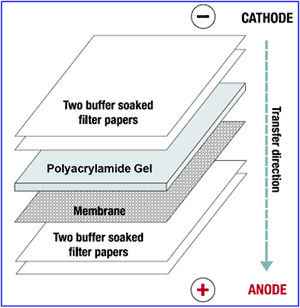
Membrane Arrangement: filter papers on top, gel, western blot membrane, filter papers at the bottom.
Image Source : kollewin.com
Transfer & Staining
Electric current is applied to transfer the protein from the gel to the membrane, transfer time, applied current and voltage need to be optimized to get better results. The blotting membrane can be stained using Ponceau S which is a reversible stain and the dye can be easily removed by washing the membrane with water. Staining with Ponceau S is helpful to know the effectiveness of protein transfer and which doesn't have any deleterious effect on the protein, if the protein is not transferred to the membrane further processing will be waste of time.
Blocking
The membrane supports used in Western blotting have a high affinity for proteins. Therefore, after the transfer of the proteins from the gel, it is important to block the remaining surface of the membrane to prevent nonspecific binding of the detection antibodies during subsequent steps. Many Blocking buffers are available to block the free sites on the membrane, BSA, non fat dry milk powder etc in PBS(phosphate buffered saline) or TBS (Tris buffered saline) with minute percentage of tween 20 or Triton X-100 can be used as blocking buffer. blocking is done overnight at 4oC. The protein in the blocking solution will attach to the membrane in all places where the target proteins have not attached. thus when antibody is added which will bind only to the target protein so background interference will be reduced.
Incubation with Primary & Secondary Antibodies
After blocking the membrane overnight, excess blocking agent is removed by washing the membrane with PBS and tween 20 (0.1%) (wash buffer) for sometime, then membrane is incubated in 1o Antibody solution (antibody solution is made in PBS and Tween 20 (0.1%), appropriate antibody dilutions need to be made which will better results, dilutions ranging from 1/50-1/500,000 can be made according to the antibody stock concentration.primary antibodies are not directly detected, tagged secondary antibodies are used for the detection.secondary antibodies are produced after detecting the antibodies belonging to a foreign species within the blood stream of vertebrate Eg: if mouse monoclonal antibodies are used as primary antibody then secondary antibody will be anti mouse IgG obtained from non-mouse host. During incubation with antibody solutions membrane is kept in gel rocker with gentle rocking. 1o antibody incubation is done for 30 mins to 2hrs sometimes more incubation periods are required. Membrane is washed with washing buffer for 5 mins to remove the excess primary antibodies. Similarly membrane is incubated with 2o antibody with suitable dilutions. incubation period varies from 30 mins to 2 hrs, secondary antibody will bind to the primary antibody, secondary antibodies are conjugated with enzyme which on reaction with substrate in the developing solution will yield colour. excess antibodies are washed off with wash buffer. there are variety of tags or enzyme labels can be conjugated to the secondary antibody. Most widely used enzymes are Horse Radish Peroxidase(HRP) and Alkaline Phosphatase (AP).sometimes radioisotopes fluorophores etc are also used, radioisotopes are expensive and has short half-life period.
Developing
Developing an immunoblot of HRP conjugated antibody is explained here, developing solution is made by dissolving the substrate 0.03% diaminobenzidene(DAB) in PBS and 1% CoCl2, to this 0.1% hydrogen peroxide (H2O2)is added prior to the incubation with the membrane. even preformulated DAB solutions are readily available. Wash the membrane with wash buffer and incubate with developing solution for 30 seconds to one minute, DAB reacts with HRP in the presence of peroxide to yield an insoluble brown-colored product at locations where peroxidase-conjugated antibodies are bound to the target protein. The reaction can be stopped by adding water.
Western Blot Membrane Showing Protein Bands
There are various other methods to develop the blot depending on the nature of tag or label used in the secondary antibody.
Important: Touching the membrane with bare hands will give background while developing. Rocking the membrane for sometime with the blocking buffer is shown to give better results. Appropriate dilutions of antibody will yield good results.
Application in Medical Diagnostics
Application in Medical Diagnostics
- HIV western blot test is used as confirmatory test for detecting anti-HIV antibody in human serum sample
- Western Blotting is also used for confirmatory test for Hepatitis B Infection.
- Testing Lyme disease
References
http://www.millipore.com/immunodetection/id3/membraneselection
http://www.bio.davidson.edu/courses/genomics/method/Westernblot.html
http://www.piercenet.com/browse.cfm?fldID=8259A7B6-7DA6-41CF-9D55-AA6C14F31193#blockingnonspecificsites
http://www.piercenet.com/browse.cfm?fldID=01041015
http://www.gelifesciences.com/aptrix/upp01077.nsf/content/ecl~Amersham_ECL_prime/$file/28982942AA.pdf


Hello,
ReplyDeleteThank you for the good writeup. Secondary antibodies are formed by the immune systems of a vertebrate upon detection of antibodies belonging to a foreign species within the blood stream, which is used to detect an unconjugated primary antibody that has bound to a target antigen...
Thank you for the additional information provided...I will update it..
ReplyDelete