What is Dot Blot?
Dot Blot is a cheaper, easier and faster technique to detect the presence of Proteins and Nucleic Acids in a biological sample. Due to the simplicity of the technique it widely used as a ideal diagnostic tool.
This post covers the Protein Dot Blot Technique.
Dot Blot Principle
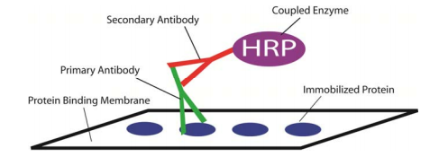
Dot Blot works based on the immunodetection principle for identifying specific protein.
Dot Blot - Technique: Overview
Proteins are immobilized on to a protein binding membrane, PVDF or Nitrocellulose. After Protein Immobilization membrane is blocked to avoid non-specific binding, using blocking buffer.
Immobilized protein is then incubated with a primary antibody (for 30mins to 1 hour). The antibody is specific to the protein and is visualized either with a specific tag or with a secondary antibody.
The secondary antibody generally will carry a tag that allow the visualization of protein.
Blots can be visualized using methods such as Chemiluminescence or Fluorescence.
Common Tags used in Dot Blots are enzymes conjugated to the secondary antibody. Upon addition of substrate, enzyme acts on the substrate to develop colour. Commonly preferred enzymes are HRP(Horse Radish Peroxidase) and Alkaline Phosphatase (AP)
Advantages of Dot Blot over Western Blot
Non-Electrophoresis Technique:
Dot Blot is non-electrophoresis technique, means that one need not do protein electrophoresis. Proteins can directly immobilized on to the membrane.
Simple & Easy Method:
No messy work of PAGE run and gel to membrane transfers, proteins are directly immobilized on membrane, making this method much easier and faster.
Time:
It is a faster method, takes ~3hours, whereas western blot may even take up to 2 days. Including the protein electrophoresis and gel to membrane transfer optimization.
Dot Blot is not as informative as Western Blot, but it is inexpesive, quick and easy method for the simple protein detection.
Dot Blot is a cheaper, easier and faster technique to detect the presence of Proteins and Nucleic Acids in a biological sample. Due to the simplicity of the technique it widely used as a ideal diagnostic tool.
This post covers the Protein Dot Blot Technique.
Dot Blot Principle
Dot Blot works based on the immunodetection principle for identifying specific protein.
 |
| Dot Blot - Principle |
Proteins are immobilized on to a protein binding membrane, PVDF or Nitrocellulose. After Protein Immobilization membrane is blocked to avoid non-specific binding, using blocking buffer.
Immobilized protein is then incubated with a primary antibody (for 30mins to 1 hour). The antibody is specific to the protein and is visualized either with a specific tag or with a secondary antibody.
The secondary antibody generally will carry a tag that allow the visualization of protein.
Blots can be visualized using methods such as Chemiluminescence or Fluorescence.
Common Tags used in Dot Blots are enzymes conjugated to the secondary antibody. Upon addition of substrate, enzyme acts on the substrate to develop colour. Commonly preferred enzymes are HRP(Horse Radish Peroxidase) and Alkaline Phosphatase (AP)
Advantages of Dot Blot over Western Blot
Non-Electrophoresis Technique:
Dot Blot is non-electrophoresis technique, means that one need not do protein electrophoresis. Proteins can directly immobilized on to the membrane.
Simple & Easy Method:
No messy work of PAGE run and gel to membrane transfers, proteins are directly immobilized on membrane, making this method much easier and faster.
Time:
It is a faster method, takes ~3hours, whereas western blot may even take up to 2 days. Including the protein electrophoresis and gel to membrane transfer optimization.
Dot Blot is not as informative as Western Blot, but it is inexpesive, quick and easy method for the simple protein detection.
No comments:
Post a Comment